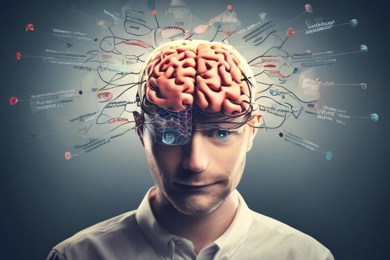
Integrating Neuroimaging Techniques in Psychological Research
In the ever-evolving landscape of psychological research, the integration of neuroimaging techniques has emerged as a game changer. Imagine being able to peek inside the human brain while it processes thoughts, emotions, and memories. This is not just science fiction; it's the reality that neuroimaging offers. By utilizing advanced imaging methods, researchers can visualize brain activity and structure, paving the way for a deeper understanding of mental processes and disorders.
Neuroimaging encompasses a variety of methods, including Functional Magnetic Resonance Imaging (fMRI), Positron Emission Tomography (PET), and Electroencephalography (EEG). Each of these techniques offers unique insights into how our brains function and respond to different stimuli. For instance, fMRI allows scientists to observe real-time brain activity by measuring changes in blood flow, while EEG captures electrical activity through electrodes placed on the scalp. These methods are not just technical marvels; they have profound implications for understanding psychological phenomena.
As we delve deeper into the applications of neuroimaging within cognitive psychology, we uncover a treasure trove of insights into how we think, remember, and make decisions. This intersection of neuroscience and psychology is crucial for unraveling the complexities of the human mind. With neuroimaging, researchers can explore intricate processes such as memory encoding and retrieval, shedding light on how we store and recall information. For example, studies utilizing fMRI have pinpointed specific brain regions activated during memory tasks, revealing the neural networks that underpin our ability to remember.
Moreover, neuroimaging techniques are instrumental in understanding emotional processing. By examining brain activity in response to emotional stimuli, researchers can identify patterns that correlate with mood disorders like depression and anxiety. This knowledge not only enhances our understanding of these conditions but also informs the development of targeted therapeutic interventions. As we continue to explore the rich tapestry of the human brain through neuroimaging, we unlock new pathways for treatment and recovery.
In summary, the integration of neuroimaging techniques in psychological research is not just a trend; it represents a significant leap forward in our quest to understand the human mind. By visualizing brain activity and correlating it with psychological processes, researchers are better equipped to tackle mental health challenges, paving the way for more effective treatments and interventions. The future of psychological research is bright, and neuroimaging is leading the charge.
- What are the main types of neuroimaging techniques? The primary neuroimaging techniques include fMRI, PET, and EEG, each serving different purposes in research.
- How does neuroimaging help in understanding mental disorders? Neuroimaging allows researchers to identify brain activity patterns associated with various disorders, facilitating early diagnosis and tailored treatments.
- Can neuroimaging predict treatment outcomes? Yes, neuroimaging can measure changes in brain activity before and after treatment, helping to evaluate the effectiveness of therapeutic interventions.

Understanding Neuroimaging Techniques
Neuroimaging is an exciting field that combines psychology and advanced technology to explore the complexities of the human brain. Imagine being able to see the brain light up like a Christmas tree, revealing the intricate dance of neurons as they communicate during various mental processes. This visualization is made possible through several neuroimaging techniques, each with its unique strengths and applications in psychological research. The most prominent methods include Functional Magnetic Resonance Imaging (fMRI), Positron Emission Tomography (PET), and Electroencephalography (EEG). Each of these techniques offers a different perspective on brain activity, allowing researchers to peel back the layers of the mind and gain insights into how we think, feel, and behave.
Functional Magnetic Resonance Imaging (fMRI) is one of the most widely used neuroimaging techniques in psychology. It tracks blood flow in the brain, providing a real-time map of brain activity. When a specific area of the brain is more active, it requires more oxygen, leading to increased blood flow. This technique allows researchers to pinpoint which regions are involved in tasks like memory recall or emotional processing. For instance, studies utilizing fMRI have shown that the hippocampus is crucial for memory formation, while the amygdala plays a vital role in emotional responses.
Positron Emission Tomography (PET), on the other hand, involves injecting a radioactive tracer into the bloodstream. This tracer binds to glucose, which brain cells use for energy. As a result, PET scans can visualize metabolic activity in different brain regions. While PET is less commonly used than fMRI due to its higher cost and lower resolution, it has been instrumental in studying various psychological disorders, such as depression and schizophrenia, by highlighting areas of the brain that are underactive or overactive.
Lastly, Electroencephalography (EEG) measures electrical activity in the brain using sensors placed on the scalp. This technique offers excellent temporal resolution, meaning it can detect changes in brain activity within milliseconds. EEG is particularly useful in cognitive psychology, where understanding the timing of brain processes is crucial. For example, researchers can use EEG to investigate how quickly the brain responds to emotional stimuli, providing insights into the mechanisms underlying anxiety and stress.
Each of these neuroimaging techniques has its advantages and limitations, making them suitable for different research questions. For instance, while fMRI provides detailed images of brain structures and functions, it cannot capture rapid changes in brain activity as effectively as EEG. Conversely, while EEG excels in temporal resolution, it does not offer the spatial precision of fMRI. Thus, researchers often combine these techniques to gain a more comprehensive understanding of mental processes.
In summary, neuroimaging techniques have revolutionized psychological research by offering a window into the brain's inner workings. By leveraging these advanced methods, scientists can unravel the complexities of cognition, emotion, and behavior, ultimately leading to better diagnostic tools and treatment options for mental health disorders. As we continue to explore the intersection of neuroimaging and psychology, the potential for new discoveries and innovations is virtually limitless.

Applications in Cognitive Psychology
Neuroimaging has transformed the landscape of cognitive psychology, offering unprecedented insights into the complex workings of the human mind. By employing advanced imaging techniques, researchers can visualize brain activity in real-time, allowing them to unravel the intricate processes underlying cognition. Imagine being able to peek inside someone’s head as they solve a puzzle or recall a cherished memory; that’s the power of neuroimaging! With methods like fMRI (functional Magnetic Resonance Imaging) and EEG (Electroencephalography), psychologists can observe how different areas of the brain light up during various cognitive tasks.
One of the most fascinating applications of neuroimaging in cognitive psychology is its role in studying memory. Through neuroimaging, researchers have been able to pinpoint specific brain regions involved in encoding, storage, and retrieval of memories. For instance, studies have shown that the hippocampus plays a crucial role in forming new memories, while the prefrontal cortex is essential for retrieving them. This understanding not only enhances our knowledge of memory processes but also has significant implications for addressing memory-related disorders.
Additionally, neuroimaging techniques allow researchers to explore the cognitive processes of attention and decision-making. By analyzing brain activity during tasks that require focused attention, scientists have identified neural pathways that contribute to how we concentrate and filter out distractions. In decision-making scenarios, neuroimaging helps to reveal how emotions and cognitive evaluations interact, shedding light on why we make certain choices over others. This dual insight into both emotional and cognitive factors is a game-changer for understanding human behavior.
To illustrate the impact of neuroimaging in cognitive psychology, consider the following table summarizing key findings from recent studies:
| Study | Technique Used | Key Findings |
|---|---|---|
| Memory Encoding | fMRI | Identified the hippocampus as critical for new memory formation. |
| Attention Focus | EEG | Revealed neural pathways involved in filtering distractions. |
| Decision-Making | fMRI | Showed interaction between emotional responses and cognitive evaluations. |
In summary, the integration of neuroimaging in cognitive psychology is not just a trend; it’s a revolution. By providing a window into the brain’s activity, these techniques have opened up new avenues for understanding how we think, remember, and make decisions. As researchers continue to explore the depths of cognitive processes through neuroimaging, we can expect exciting advancements that will further enrich our comprehension of the human mind.
- What is neuroimaging? Neuroimaging refers to a range of techniques used to visualize brain activity and structure, including fMRI, PET, and EEG.
- How does neuroimaging help in cognitive psychology? It allows researchers to observe brain activity associated with cognitive processes such as memory, attention, and decision-making.
- What are the implications of neuroimaging findings? Discoveries from neuroimaging can lead to better understanding and treatment of cognitive disorders, enhancing therapeutic approaches.

Memory and Neuroimaging
Memory is one of the most fascinating aspects of human cognition, and thanks to neuroimaging, we can now peer into the intricate workings of the brain as it encodes, stores, and retrieves information. Imagine your brain as a vast library, where every experience, thought, and emotion is cataloged in its own section. Neuroimaging techniques like fMRI (functional Magnetic Resonance Imaging) and PET (Positron Emission Tomography) allow researchers to see which parts of this library are active when we remember something. This not only enhances our understanding of memory but also opens doors to new treatments for memory-related disorders.
Through the lens of neuroimaging, we can identify specific brain regions that play crucial roles in memory processes. For instance, studies have shown that the hippocampus is vital for forming new memories, while the prefrontal cortex is key in retrieving them. By observing brain activity in real-time, researchers have been able to map out the journey of a memory from the moment it is created to when it is recalled, revealing a dynamic interplay between various brain regions. This understanding is pivotal, especially when we consider how memory can be affected by age, trauma, and mental health conditions.
Let’s break down the memory process into three main stages, akin to the lifecycle of a book in our library analogy:
- Encoding: This is the initial stage where information is transformed into a format that can be stored. Neuroimaging studies reveal that during this phase, the brain's hippocampus and surrounding areas are particularly active.
- Storage: Once information is encoded, it needs to be stored for future retrieval. Here, various brain regions work together to consolidate memories, often involving the neocortex.
- Retrieval: This is the process of accessing stored information. Neuroimaging has shown that different brain areas, including the prefrontal cortex, become activated during recall, showcasing how complex and interconnected our memory systems are.
Research has highlighted numerous key studies that have advanced our understanding of these memory mechanisms. For example, one groundbreaking study used fMRI to track brain activity while participants engaged in memory tasks. The results showed distinct patterns of activation corresponding to successful memory recall versus failed attempts. Such insights are invaluable, not just for academic purposes but also for practical applications in education and therapy.
Moreover, neuroimaging has profound implications for understanding memory-related disorders, such as Alzheimer’s disease. By identifying early changes in brain activity, researchers can develop targeted interventions that may slow the progression of these conditions. It's like having a roadmap that guides us through the complexities of memory, helping us navigate challenges that arise as we age or face cognitive decline.
In summary, the integration of neuroimaging in memory research is revolutionizing our comprehension of how memories are formed, stored, and recalled. As we continue to explore this uncharted territory, the potential for new therapies and educational strategies grows, promising a brighter future for those struggling with memory issues. The more we understand about the brain's memory systems, the more tools we have at our disposal to enhance cognitive health and resilience.
- What is neuroimaging? Neuroimaging refers to a variety of techniques used to visualize the structure and function of the brain, including fMRI, PET, and EEG.
- How does neuroimaging help in understanding memory? It allows researchers to observe which brain areas are active during different memory processes, providing insights into how memories are formed, stored, and retrieved.
- Can neuroimaging assist in treating memory disorders? Yes! By identifying changes in brain activity, neuroimaging can help develop targeted therapies for conditions like Alzheimer’s disease.

Encoding Processes
Understanding how information is encoded in the brain is vital for cognitive psychology, as it lays the groundwork for how we learn and remember. Neuroimaging studies have significantly advanced our knowledge of these encoding processes, revealing how different brain regions collaborate to transform sensory input into lasting memories. For instance, functional Magnetic Resonance Imaging (fMRI) allows researchers to observe which areas of the brain are activated when individuals are exposed to new information. This has led to some fascinating insights into how the brain organizes and stores memories.
During the encoding phase, the brain engages in a complex dance of electrical and chemical signals. Various regions, such as the hippocampus, prefrontal cortex, and parietal lobes, play crucial roles in this process. The hippocampus is often referred to as the brain's memory center, where initial encoding occurs. Meanwhile, the prefrontal cortex is involved in the integration and manipulation of information, ensuring that what we learn can be efficiently stored and retrieved later. To illustrate this, consider the following table that summarizes key brain regions involved in memory encoding:
| Brain Region | Function in Encoding |
|---|---|
| Hippocampus | Initial encoding of new information |
| Prefrontal Cortex | Integration and manipulation of information |
| Parietal Lobes | Spatial awareness and sensory integration |
Moreover, neuroimaging has uncovered the significance of attention in the encoding process. When we focus intently on a task or piece of information, our brain's encoding efficiency increases. This is where the interplay between attention and memory becomes crucial. For example, studies have shown that when participants are asked to memorize a list of words while being distracted, their brain activity differs significantly compared to when they are fully engaged. This leads us to ponder: how can we harness this knowledge to improve our learning strategies?
One intriguing finding is that the emotional context of the information being encoded can also enhance memory retention. Research indicates that emotionally charged experiences are more likely to be remembered due to heightened activity in the amygdala, which interacts with the hippocampus during encoding. This connection between emotion and memory suggests that if we want to improve our memory, we might consider adding an emotional component to our learning experiences. After all, who hasn’t remembered a moment from their life because it was tied to a powerful emotion?
In summary, neuroimaging techniques provide invaluable insights into the encoding processes of memory. By revealing the intricate workings of the brain during this phase, researchers can better understand how we learn and retain information. This knowledge not only enriches the field of cognitive psychology but also opens up exciting possibilities for enhancing educational methods and therapeutic interventions.
- What is neuroimaging? Neuroimaging refers to a variety of techniques used to visualize brain activity and structure, helping researchers understand how the brain works.
- How does fMRI work? Functional Magnetic Resonance Imaging (fMRI) measures brain activity by detecting changes in blood flow, allowing researchers to see which areas are active during specific tasks.
- Why is encoding important in memory? Encoding is the process by which information is transformed into a format that can be stored in memory, making it essential for learning and recall.
- Can emotions affect memory encoding? Yes, emotional experiences can enhance memory encoding, making it more likely that we will remember emotionally charged information.

Retrieval Mechanisms
When we think about memory, it's not just a static filing cabinet; it’s more like a dynamic web of connections that we navigate every day. The process of retrieval is crucial in understanding how we access stored information, and neuroimaging has opened a window into the brain's inner workings during this fascinating process. Through various imaging techniques, researchers can observe which brain regions light up when we recall a memory, shedding light on the intricate dance of neurons that occurs behind the scenes.
One of the most significant findings in neuroimaging studies is the activation of specific brain areas during memory retrieval. For instance, the hippocampus often shows heightened activity when we are trying to recall past experiences. This small, seahorse-shaped structure is vital for forming new memories and plays a key role in retrieving them as well. Additionally, the prefrontal cortex, which is involved in higher cognitive functions such as decision-making and problem-solving, also lights up during recall tasks. This suggests that our brain doesn't just pull memories from a shelf; it actively reconstructs them, often influenced by context and current emotional states.
Moreover, neuroimaging studies have revealed that the strength of neural connections can impact the efficiency of memory retrieval. For example, individuals with stronger connections in the default mode network—a network of brain regions that are active when we are at rest and not focused on the external environment—tend to exhibit better recall abilities. This highlights the idea that memory is not merely about what we have stored but also about how well we can access that information when needed.
Interestingly, neuroimaging has also provided insights into the phenomenon known as the tip-of-the-tongue (TOT) state, where we know we know something but can’t quite access it. Studies indicate that during these moments, there is often partial activation of the relevant brain regions, suggesting that the information is close to being retrieved but not quite there yet. This has profound implications for therapeutic approaches, as understanding these mechanisms can help in developing strategies to assist individuals with memory retrieval difficulties, particularly in conditions like Alzheimer’s disease.
In summary, the exploration of retrieval mechanisms through neuroimaging not only enhances our understanding of how memories are accessed but also opens up new avenues for therapeutic interventions. By pinpointing the brain regions involved, we can better tailor treatments for individuals struggling with memory-related issues, making neuroimaging an invaluable tool in both psychological research and clinical practice.
- What is neuroimaging? Neuroimaging refers to a variety of techniques used to visualize the structure and function of the brain. Common methods include fMRI, PET, and EEG.
- How does neuroimaging help in psychological research? It allows researchers to observe brain activity in real-time, providing insights into cognitive processes, emotional responses, and the effects of psychological disorders.
- What are the implications of understanding retrieval mechanisms? By understanding how memories are retrieved, researchers can develop better therapeutic strategies for individuals with memory impairments or disorders.
- Can neuroimaging be used in clinical settings? Absolutely! Neuroimaging can aid in diagnosing mental health disorders and evaluating the effectiveness of treatments.

Emotional Processing and Neuroimaging
Neuroimaging techniques have revolutionized our understanding of how emotions are processed in the brain, offering a window into the intricate dance of neural activity that accompanies our feelings. By utilizing advanced imaging methods like functional Magnetic Resonance Imaging (fMRI) and Electroencephalography (EEG), researchers can observe real-time brain responses to emotional stimuli. Imagine being able to see the brain light up in response to joy, fear, or sadness—it's like watching a fireworks display of activity that reveals the underlying mechanisms of our emotional lives.
One of the key insights gained from neuroimaging studies is the identification of specific brain regions that are activated during emotional experiences. For instance, the amygdala, a small almond-shaped structure, is crucial for processing fear and other emotions. When we encounter a threatening situation, the amygdala springs into action, triggering a cascade of physiological responses. This is just one example of how neuroimaging helps us map the emotional landscape of the brain.
Furthermore, neuroimaging allows researchers to explore how individual differences in brain activity relate to emotional responses. For example, some people may show heightened amygdala activity when exposed to fearful stimuli, indicating a predisposition to anxiety disorders. This understanding can pave the way for personalized therapeutic approaches, where treatments are tailored based on an individual's unique neural profile.
Moreover, the implications of these findings extend beyond basic research. By analyzing brain activity patterns associated with various emotions, clinicians can develop targeted interventions for mood disorders. For instance, understanding how the brain processes positive emotions can inform therapies aimed at enhancing emotional well-being in individuals suffering from depression.
In summary, the integration of neuroimaging in emotional processing research offers profound insights into the neural correlates of our feelings. As we continue to unravel the complexities of the emotional brain, we move closer to developing effective treatments that address the root causes of emotional disturbances, ultimately improving mental health outcomes for many.
- What is neuroimaging? Neuroimaging refers to a variety of techniques used to visualize brain activity and structure, helping researchers understand mental processes and disorders.
- How does neuroimaging contribute to emotional processing research? It allows researchers to observe real-time brain activity in response to emotional stimuli, identifying specific brain regions involved in processing emotions.
- What are some common neuroimaging techniques? Common techniques include functional Magnetic Resonance Imaging (fMRI), Positron Emission Tomography (PET), and Electroencephalography (EEG).
- Can neuroimaging help in treating emotional disorders? Yes, by identifying brain activity patterns associated with emotional responses, neuroimaging can inform personalized treatment approaches for mood disorders.

Neuroimaging in Clinical Psychology
Neuroimaging techniques have revolutionized the field of clinical psychology, providing a window into the intricate workings of the human brain. By visualizing brain activity and structure, researchers and clinicians can better understand mental disorders and develop more effective treatment strategies. Imagine being able to see the brain in action, pinpointing exactly where things might be going awry. This is the power of neuroimaging!
One of the most significant contributions of neuroimaging in clinical psychology is its ability to enhance diagnostic accuracy. Traditional methods of diagnosing mental disorders often rely on subjective reports and behavioral assessments, which can sometimes be misleading. In contrast, neuroimaging offers objective data that can reveal underlying neurological issues. For instance, studies using functional Magnetic Resonance Imaging (fMRI) have shown distinct patterns of brain activity associated with conditions like depression, anxiety, and schizophrenia. This means that clinicians can make more informed decisions and tailor their approach to individual patients.
Moreover, neuroimaging is pivotal in identifying biomarkers for various psychological disorders. A biomarker is a biological indicator that can signal the presence of a disease or condition. By utilizing techniques such as Positron Emission Tomography (PET) and Electroencephalography (EEG), researchers have begun to uncover specific brain signatures linked to disorders like ADHD and PTSD. These findings are not just academic; they hold the potential for early diagnosis and personalized treatment options that can significantly improve patient outcomes.
In addition to diagnosis, neuroimaging plays a crucial role in evaluating treatment efficacy. As therapies evolve, whether they be pharmacological or psychotherapeutic, it's essential to measure their impact on the brain. Neuroimaging allows clinicians to observe changes in brain activity following interventions. For example, studies have shown that cognitive-behavioral therapy (CBT) can lead to observable changes in brain function in individuals with anxiety disorders. By comparing brain scans before and after treatment, clinicians can assess how well a patient is responding to therapy and make necessary adjustments.
Furthermore, the integration of neuroimaging into clinical practice opens up exciting possibilities for the future. As technology advances, we may see even more sophisticated imaging techniques that can provide deeper insights into the brain's functioning. Imagine a world where mental health treatments are as personalized as a tailored suit, where each intervention is based on a patient's unique brain profile. This is not just a dream; it's becoming a reality, thanks to neuroimaging.
In conclusion, the integration of neuroimaging into clinical psychology is not just a trend; it is a transformative approach that enhances our understanding of mental disorders and improves treatment strategies. As we continue to explore the complexities of the brain, we are likely to uncover even more groundbreaking insights that will shape the future of psychological research and practice.
- What is neuroimaging? Neuroimaging refers to a variety of techniques used to visualize the structure and function of the brain.
- How does neuroimaging help in clinical psychology? It provides objective data for diagnosing mental disorders, identifies biomarkers, and evaluates treatment efficacy.
- What are some common neuroimaging techniques? Common techniques include fMRI, PET, and EEG.
- Can neuroimaging predict treatment outcomes? Yes, by assessing brain activity changes, neuroimaging can help predict how well a patient may respond to a specific treatment.

Identifying Biomarkers for Disorders
Neuroimaging is revolutionizing the way we understand psychological disorders by enabling researchers to identify biomarkers associated with various mental health conditions. A biomarker, in this context, is a measurable indicator of a biological state or condition, which can help in diagnosing disorders, predicting their progression, and tailoring personalized treatment plans. For instance, through advanced imaging techniques like fMRI (functional Magnetic Resonance Imaging) and PET (Positron Emission Tomography), scientists can observe the brain's activity and structure, providing insights into how different regions may function abnormally in individuals with psychological disorders.
One of the most compelling aspects of neuroimaging is its ability to reveal patterns of brain activity that correlate with specific disorders. For example, studies have shown that individuals with depression often exhibit reduced activity in the prefrontal cortex, a region associated with decision-making and emotional regulation. Similarly, neuroimaging has identified hyperactivity in the amygdala among those with anxiety disorders, highlighting its role in processing fear and stress. These findings are not just academic; they carry significant implications for clinical practice, offering pathways for early diagnosis and intervention.
Moreover, the identification of biomarkers through neuroimaging can lead to the development of targeted therapies. For instance, if a specific brain pattern is associated with a disorder, clinicians can tailor treatment strategies that specifically address those neural mechanisms. This could be particularly beneficial in the realm of psychopharmacology, where medications can be prescribed based on the unique neurobiological profile of the patient. By aligning treatments with the underlying brain activity, the efficacy of interventions can be significantly enhanced.
As we delve deeper into the world of neuroimaging, it’s essential to recognize its limitations as well. While the identification of biomarkers is promising, it is only one piece of the puzzle. Psychological disorders are complex and multifaceted, influenced by a myriad of factors including genetics, environment, and personal history. Therefore, while neuroimaging provides invaluable insights, it should be integrated with other diagnostic tools and clinical assessments to form a comprehensive understanding of a patient's condition.
In summary, the integration of neuroimaging in identifying biomarkers for psychological disorders is a game-changer. It not only enhances our understanding of the biological underpinnings of mental health issues but also paves the way for more effective and personalized treatment approaches. As research continues to evolve, we can anticipate a future where mental health care is as precise and targeted as other fields of medicine.
- What are biomarkers in psychology? Biomarkers in psychology are measurable indicators of biological states or conditions that can assist in diagnosing and treating mental health disorders.
- How does neuroimaging help in identifying disorders? Neuroimaging allows researchers to observe brain activity and structure, revealing patterns that correlate with specific psychological disorders.
- Can neuroimaging lead to personalized treatment? Yes, by identifying unique brain activity patterns associated with disorders, neuroimaging can inform targeted therapy strategies for individual patients.
- What are some limitations of neuroimaging? While neuroimaging provides valuable insights, psychological disorders are complex and influenced by various factors, necessitating a comprehensive approach to diagnosis and treatment.

Evaluating Treatment Efficacy
In the ever-evolving landscape of psychology, the need to evaluate the efficacy of treatments is paramount. With the integration of neuroimaging techniques, researchers are now equipped with the tools to observe the brain's response to various therapeutic interventions in real-time. This advancement has revolutionized how we understand the effectiveness of treatments for mental health disorders. Imagine being able to see the brain's activity light up like a city skyline at night, revealing which areas are activated during therapy sessions. This is the power of neuroimaging!
One of the most significant benefits of using neuroimaging in evaluating treatment efficacy is its ability to provide objective data. Traditional methods of assessing treatment success often rely on self-reported measures, which can be subjective and influenced by a myriad of factors, including mood and perception. However, neuroimaging allows researchers to visualize changes in brain activity, offering a more concrete understanding of how treatments are affecting individuals. For instance, studies using functional Magnetic Resonance Imaging (fMRI) have shown how brain patterns change in response to cognitive-behavioral therapy (CBT) in patients with anxiety disorders.
Moreover, neuroimaging can help identify which specific treatments are most effective for different individuals. This personalized approach is crucial because mental health disorders are complex and can manifest differently from one person to another. By analyzing brain activity patterns, clinicians can tailor interventions based on the unique neurobiological profile of each patient. For example, a patient with depression might show different neural responses to mindfulness-based therapy compared to someone with PTSD. Understanding these differences can lead to more effective and targeted treatment plans.
Additionally, neuroimaging techniques can be used to track progress over time. By conducting scans before, during, and after treatment, researchers can monitor changes in brain activity and structure. This longitudinal approach not only helps in assessing the immediate effects of therapy but also provides insights into long-term outcomes. For example, a study may reveal that certain brain regions associated with emotional regulation exhibit increased activity after a series of therapy sessions, indicating that the treatment is fostering healthier emotional processing.
As we delve deeper into the implications of neuroimaging for treatment efficacy, it's essential to consider the ethical dimensions. While the potential benefits are immense, researchers must navigate the complexities of privacy and consent when it comes to brain imaging. Patients should be informed about how their data will be used and the potential risks involved. Transparency in this process is crucial for maintaining trust between clinicians and patients.
In summary, evaluating treatment efficacy through neuroimaging is a groundbreaking approach that enhances our understanding of mental health interventions. By providing objective data, personalizing treatment plans, and tracking progress over time, neuroimaging paves the way for more effective and tailored psychological care. As research continues to unfold, we can anticipate even more innovative applications of these techniques, ultimately leading to better outcomes for individuals seeking help.
- What is neuroimaging? Neuroimaging refers to a variety of techniques used to visualize the structure and function of the brain, including fMRI, PET, and EEG.
- How does neuroimaging help in evaluating treatment efficacy? Neuroimaging provides objective data on brain activity, allowing researchers to assess how different treatments affect mental health disorders.
- Can neuroimaging personalize treatment plans? Yes, by analyzing brain activity patterns, clinicians can tailor interventions to suit the unique neurobiological profiles of their patients.
- Are there ethical concerns with neuroimaging? Yes, issues related to privacy, consent, and data usage must be carefully considered in neuroimaging research.
Frequently Asked Questions
- What are the main neuroimaging techniques used in psychological research?
Neuroimaging techniques commonly used in psychological research include functional Magnetic Resonance Imaging (fMRI), Positron Emission Tomography (PET), and Electroencephalography (EEG). Each method has its strengths, allowing researchers to visualize brain activity and structure in unique ways, providing critical insights into mental processes.
- How does neuroimaging contribute to our understanding of memory?
Neuroimaging helps researchers pinpoint specific brain regions involved in memory functions such as encoding, storage, and retrieval. By analyzing brain activity during memory tasks, scientists can uncover how information is processed and recalled, which has significant implications for enhancing learning and therapeutic strategies.
- Can neuroimaging techniques help identify psychological disorders?
Absolutely! Neuroimaging plays a crucial role in identifying biomarkers associated with various psychological disorders. These biomarkers can lead to earlier diagnoses and more personalized treatment options, improving patient outcomes significantly.
- How is neuroimaging used to evaluate treatment efficacy?
Neuroimaging allows researchers to assess how brain activity changes in response to psychological treatments. By measuring these changes, they can evaluate the effectiveness of different therapeutic interventions, providing valuable feedback for refining treatment approaches.
- What insights does neuroimaging provide about emotional processing?
Neuroimaging techniques reveal how brain activity correlates with emotional responses. This understanding is vital for comprehending mood disorders and can inform the development of targeted therapies that address specific emotional dysregulations.
- Are there any limitations to neuroimaging techniques?
Yes, while neuroimaging offers incredible insights, it also has limitations, such as high costs, the need for specialized equipment, and sometimes a lack of spatial or temporal resolution in capturing dynamic brain processes. Researchers must consider these factors when designing studies and interpreting results.